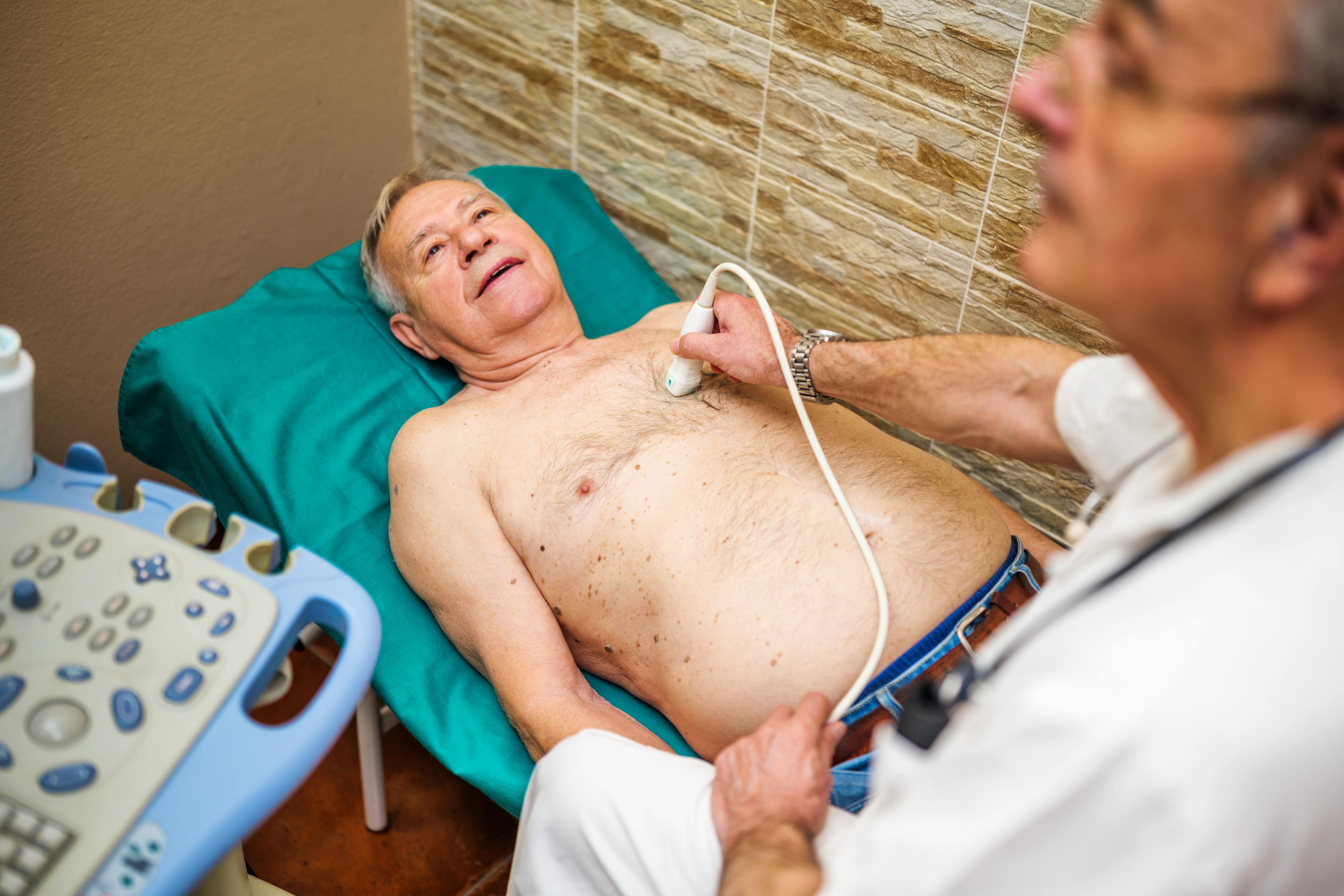

A healthy heart is critical when it comes to living a full and active lifestyle. CLS Health offers advanced echocardiography services to help diagnose and manage a wide range of heart conditions. Our expert cardiologists use cutting-edge ultrasound technology to evaluate your heart’s performance, ensuring accurate results and personalized treatment plans.
What is an Echocardiogram
An echocardiogram test uses sound waves to create real-time images of your heart. This test allows cardiologists to assess your heart's structure, pumping function, and blood flow and how efficiently it pumps blood. It is typically used to help diagnose many types of heart disease, evaluate heart failure by measuring pumping function (ejection fraction), cardiomyopathy, and valve disorders.
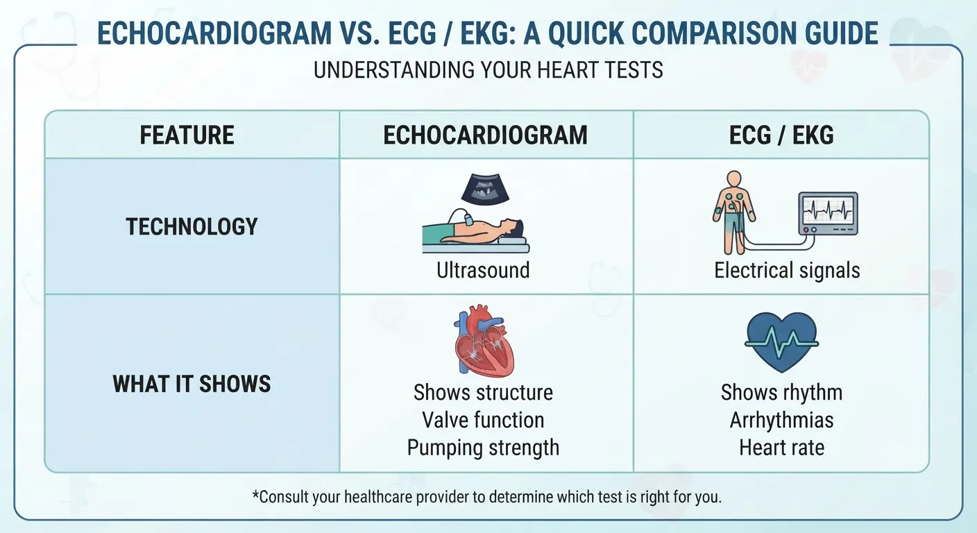
Unlike an electrocardiogram (EKG or ECG), which measures electrical activity, an echocardiogram provides a visual assessment of the heart's structure and function.
Why is an echocardiogram performed?
An echocardiogram helps evaluate your heart's structure and function. Your healthcare provider may recommend one to investigate symptoms such as chest pain, shortness of breath, palpitations, dizziness, or swelling in the legs. It may also be used to diagnose heart conditions, monitor existing heart disease, or assess heart function before or after certain treatments.
What's the difference between Echocardiogram and ECG?
What are the different types of echocardiogram?
There are different types of echocardiograms, each designed for specific diagnostic purposes. Your doctor will determine the best option based on your symptoms and medical history.
Transthoracic echocardiogram
This is the most common type of echocardiogram test. A technician places a small probe (transducer) on your chest, which sends sound waves to create images of your heart. It is painless and typically takes 30 to 60 minutes.
Transesophageal echocardiogram
If a more detailed view is needed, a transesophageal echocardiogram may be recommended. A thin, flexible tube with a transducer is gently inserted down the esophagus to capture high-resolution images. This approach is often used to assess valve function, blood clots, or congenital heart conditions.
Exercise stress echocardiogram
A stress echocardiogram evaluates how your heart responds to physical activity. Patients walk on a treadmill or take medication to increase heart rate while undergoing imaging. This test is often used to assess coronary artery disease and blood flow issues.
What happens during an echocardiogram?
During a standard transthoracic echocardiogram, you will usually lie on your back or left side. A technician applies gel to your chest and moves a small device called a transducer over the skin to create images of your heart.
The test is painless, although you may feel mild pressure from the transducer. You may be asked to change positions, breathe normally, or briefly hold your breath so the technician can capture clearer images.
How long does an echocardiogram take?
The duration of the test depends on the type of echocardiogram performed:
- Transthoracic echocardiogram: 30-60 minutes
- Transesophageal echocardiogram: 60-90 minutes (including sedation and recovery)
- Stress echocardiogram: 60 minutes (including exercise or medication administration)
The good news? Most echocardiograms require minimal preparation and allow you to return to your normal activities the same day.
When would I need an echocardiogram?
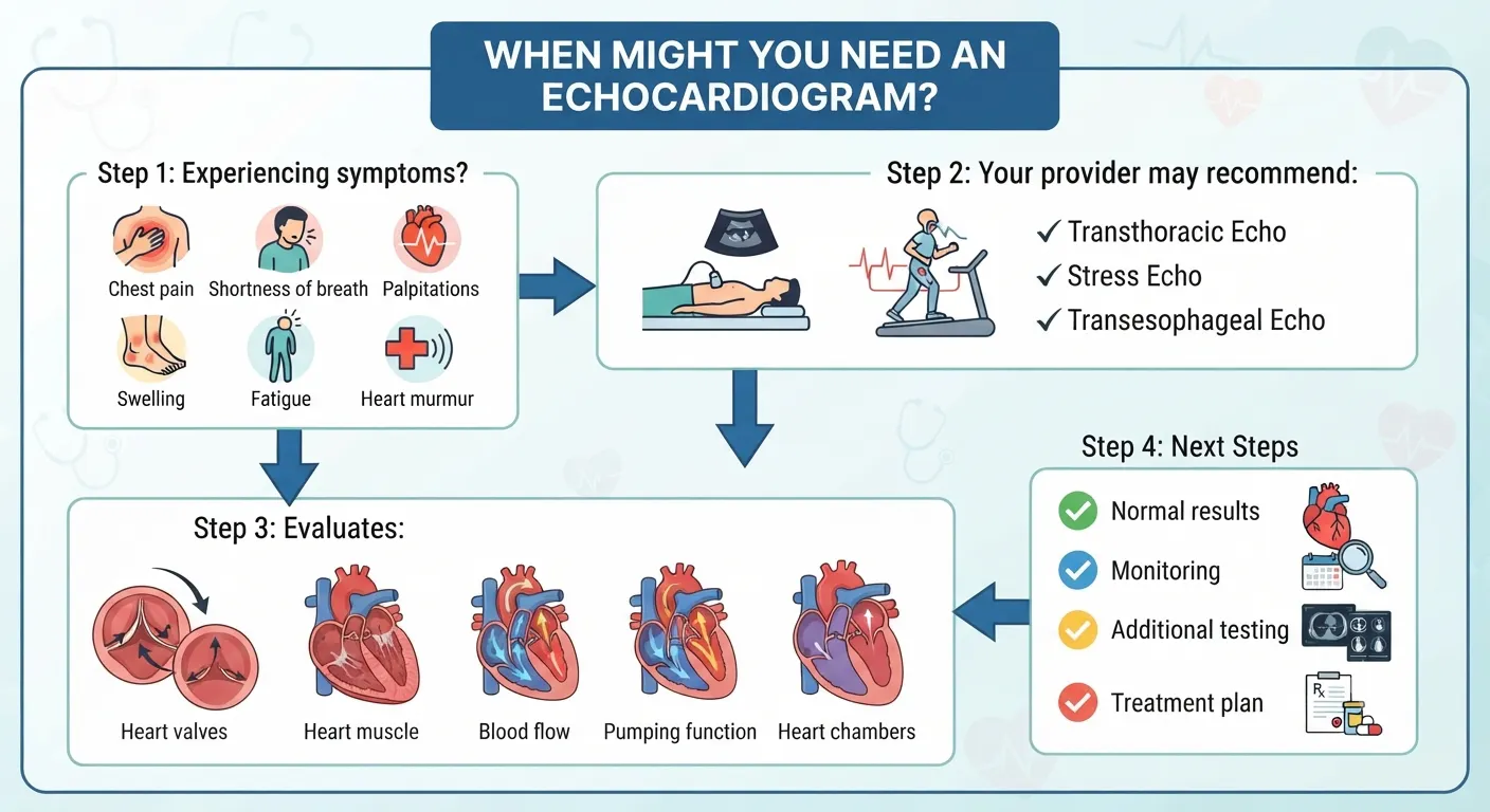
Your doctor may recommend an echocardiogram if you have:
- Chest pain
- Shortness of breath
- Unexplained fatigue
- Palpitations or an irregular heartbeat
- Dizziness or fainting
- Swelling in the legs or ankles
- Reduced ability to exercise
- A heart murmur detected during an exam
The test may also be used to monitor an existing heart condition, evaluate treatment progress, or assess heart function before or after certain procedures.
Concerned About Your Heart Symptoms?
If you're experiencing chest pain, shortness of breath, palpitations, or other symptoms that may indicate a heart condition, an echocardiogram can provide valuable information about your heart's structure and function. Our experienced cardiologists will determine whether an echocardiogram or another cardiac test is the right next step based on your symptoms and medical history.
FAQs
Preparation for an echocardiogram depends on the type of test being performed:
- Transthoracic echocardiogram (TTE): Does not require any special preparation. You can eat, drink, and take medications as usual.
- Transesophageal echocardiogram (TEE): Requires fasting for at least six hours before the test. Because sedation is used, you should arrange for someone to drive you home after the procedure.
Stress echocardiogram: May involve exercise or medication to increase heart rate. Wear comfortable clothing and avoid caffeine or certain medications as instructed by your doctor.
It depends on the type of echocardiogram. You can usually eat and drink normally before a standard transthoracic echocardiogram. A transesophageal echocardiogram generally requires fasting for several hours, while a stress echocardiogram may have separate food, caffeine, or medication instructions. Follow the preparation instructions provided by your care team.
No. A transthoracic echocardiogram is painless and non-invasive. You may feel mild pressure from the transducer. A transesophageal echocardiogram involves sedation and may cause temporary throat soreness afterward.
Your cardiologist will review your echocardiogram results and discuss the findings with you. In many cases, results are available within 24 to 72 hours, although timing may vary depending on the type of test and whether additional review is needed. Preliminary findings may be available sooner when testing is performed urgently.
Your cardiologist will explain whether your heart size, pumping function, valves, and blood flow appear normal and discuss any recommended next steps.
An electrocardiogram (EKG/ECG) records electrical activity and detects heart rhythm abnormalities. An echocardiogram, on the other hand, creates images of the heart’s structure and function, providing detailed insights into blood flow, valve function, and heart muscle health.
An echocardiogram does not directly show blocked coronary arteries, but it can identify changes in heart muscle movement or pumping function that may suggest reduced blood flow. Your cardiologist may recommend additional testing if coronary artery disease is suspected.
What does an echocardiogram diagnose?
Echocardiograms can help diagnose, evaluate, or monitor heart conditions such as:
- Heart valve disease – Identifies narrowed or leaking heart valves.
- Heart failure – Evaluates the heart’s pumping function, including ejection fraction.
- Cardiomyopathy – Detects changes in the size, thickness, or strength of the heart muscle.
- Congenital heart defects – Identifies structural heart abnormalities present from birth.
- Pericardial disease – Detects fluid buildup or other problems affecting the sac around the heart.
- Pulmonary hypertension – Helps estimate pressure in the blood vessels connecting the heart and lungs.
- Enlarged heart – Measures the size of the heart’s chambers.
- Blood clots or masses within the heart – Certain types of echocardiograms, particularly a transesophageal echocardiogram, can provide a closer view.
- Aortic abnormalities – Evaluates visible portions of the aorta, including the aortic root.
- Heart damage after a heart attack – Assesses changes in heart muscle movement and pumping function.
- Possible coronary artery disease – A stress echocardiogram may identify heart muscle changes that suggest reduced blood flow.
An echocardiogram can evaluate heart structure, function, valves, and blood flow, but it does not directly show every type of cardiovascular disease.
Schedule a Heart Evaluation in Houston
If you have symptoms such as chest pain, shortness of breath, palpitations, or your healthcare provider has recommended an echocardiogram, our experienced cardiologists are here to help. We'll evaluate your symptoms, determine whether an echocardiogram or other cardiac testing is appropriate, and create a personalized care plan.