Key Takeaways
CT scans and MRIs both create detailed internal images but use different technologies and are chosen for different clinical needs
https://providers.cls.health/search?primary_care=Primary+Care+Providers&sort=relevance%2Cnetworks%2Cavailability_density_best
Your provider selects CT vs. MRI based on the body area, symptoms, urgency, medical history, and the level of detail needed for diagnosis
Every year, millions of imaging tests are performed to help doctors diagnose injuries and medical conditions. If your doctor ordered imaging, understanding which test shows what—CT scan vs. MRI—can make the process less stressful. While both create detailed images of the inside of the body, they use different technologies and are suited for different clinical needs.
Understanding how each test works—and how imaging decisions are made—can help you feel more informed and confident about your care.
At CLS Health, imaging recommendations are coordinated through your care team. While CT imaging availability may vary by location, advanced PET/CT imaging is available when clinically appropriate.
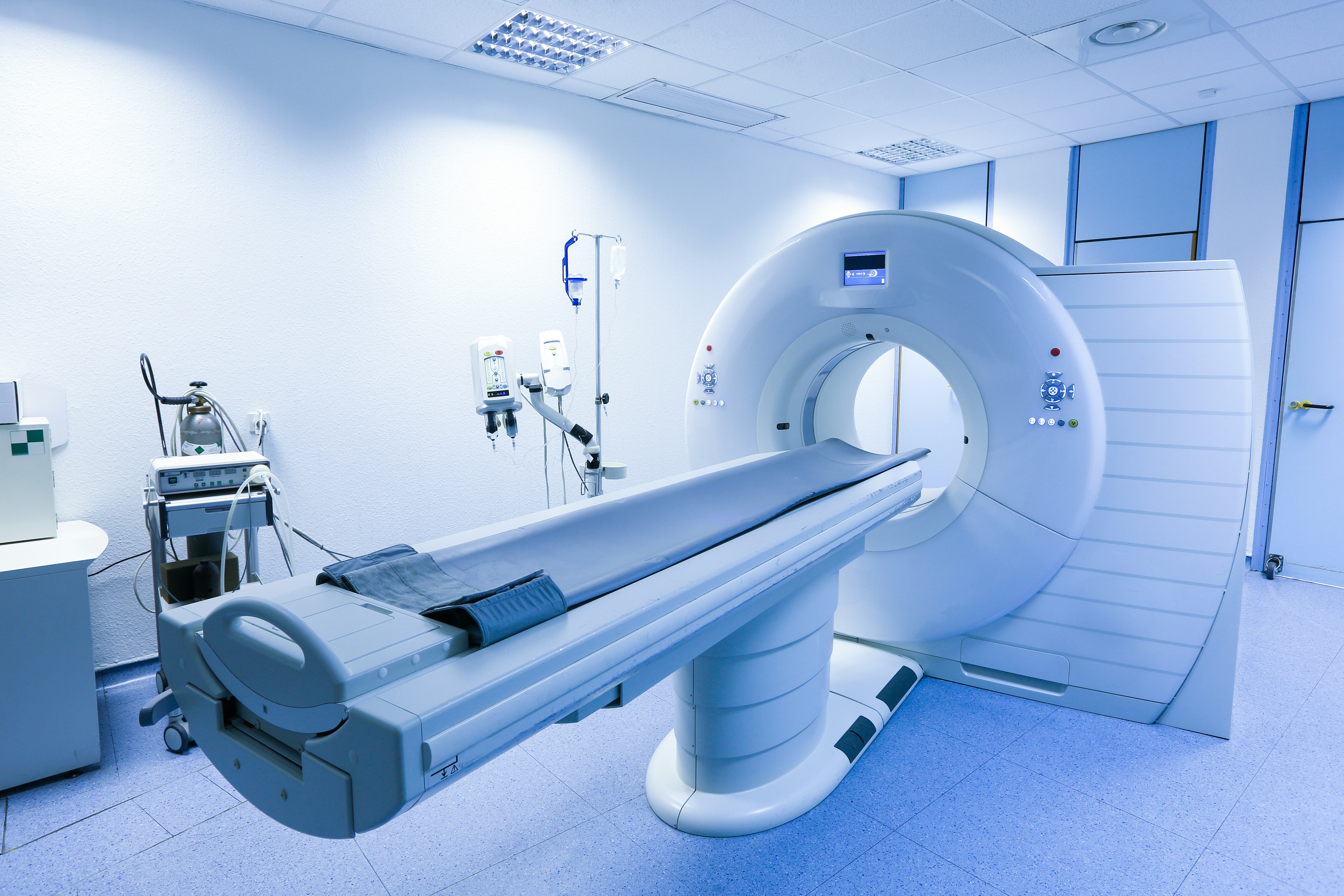
How a CT Scan Works
A CT scan (computed tomography) uses a series of X-ray images taken from multiple angles to create cross-sectional images of the body. The test is fast—often completed in just a few minutes—making it especially valuable when rapid answers are needed.
CT scans are commonly used to:
- Detect internal injuries or bleeding after trauma
- Evaluate the lungs for infection, tumors, or other abnormalities
- Assess blood vessels and circulation
- Identify and monitor certain tumors
- Guide procedures such as biopsies or surgical planning
Because CT scans use X-rays, they involve a small amount of radiation. Providers carefully weigh the benefits and risks based on a patient’s condition and medical history.
How an MRI Works
MRI (magnetic resonance imaging) uses strong magnets and radio waves instead of X-rays to create highly detailed images of soft tissues and organs. This makes MRI particularly useful when evaluating structures that may not be well visualized on CT.
MRIs are commonly used for:
- Brain and spinal cord imaging
- Joint, ligament, and muscle injuries
- Breast cancer screening in high-risk patients
- Abdominal and pelvic organ evaluation
- Detecting infections or inflammation not visible on X-ray
MRI does not involve radiation, but scans typically take longer—anywhere from 15 minutes to over an hour—and require lying still inside the scanner.
Key Differences Between CT Scans and MRIs
CT Scan Differences
- Very fast (often minutes)
- Ideal for trauma, lungs, bones, and vascular imaging
- Uses low levels of radiation
- More open scanner design
MRI Differences
- Longer exam time
- Superior detail for soft tissues, brain, and joints
- No radiation exposure
- Enclosed scanner design
Each test provides different information, and the choice depends on what your provider needs to evaluate.
How Your Provider Chooses Between CT and MRI
Your provider considers several factors when recommending imaging, including:
- The part of the body being examined
- How quickly are results needed
- Your symptoms and medical history
- Any implants, devices, or contrast allergies
- The level of detail required for diagnosis
In some cases, more than one imaging test may be used to get a complete picture.
Preparing for Your Imaging Test
CT Scan Preparation
- You may need to avoid eating or drinking if contrast is used
- Let your provider know about allergies or kidney conditions
MRI Preparation
- Remove all metal objects before the exam
- Inform your provider about implants, pacemakers, or medical devices
Your care team will provide specific instructions based on the test that has been ordered.
When PET/CT Imaging May Be Recommended
In certain situations, providers may recommend PET/CT imaging, which combines metabolic imaging with detailed anatomical imaging. PET/CT evaluates how tissues and organs are functioning—not just how they appear structurally.
PET/CT may be used for:
- Cancer diagnosis, staging, and treatment monitoring
- Evaluating cardiac viability or inflammation
- Clarifying findings from other imaging studies
This advanced imaging approach helps guide more precise diagnosis and treatment planning when clinically appropriate.
Imaging Guidance at CLS Health
Understanding imaging options helps you and your provider make informed decisions about next steps. At CLS Health, our medical imaging services—including PET/CT when indicated—are coordinated through your care team to ensure accuracy, safety, and continuity of care.
If you have questions about imaging or need help determining the right evaluation, a provider can guide you based on your symptoms and medical history.